


Contents
Scroll to:
https://doi.org/10.23947/2949-4826-2025-24-2-43-54
EDN: GPKBZO
Scroll to:
Introduction. In veterinary medicine, X-ray diagnostics is one of the most popular and affordable methods of visual diagnostics of pathologies not only of the musculoskeletal system, but also of some parenchymatous organs in the thoracic and abdominal cavities. Identification of cardiac pathologies most often begins with X-ray examination, however, the use of special techniques is sometimes required for reliable determination of the presence or absence of cardiomegaly in animals under examination. One of the most reliable methods for assessing the condition of the heart is the vertebral heart scale (VHS) method. The aim of the present review is to summarise the data available in literature sources on the use of the vertebral heart scale method during X-ray diagnostics of the feline and canine thoracic cavity, and to assess the relevance and prospects for development of this technique.
Materials and Methods. The search for the articles for the scientific review was conducted in the PubMed and eLIBRARY.RU databases. The search covered only publications in Russian and English, published between January 1, 1995 and December 31, 2024. According to the criteria of including a scientific paper in a review, the article should have met the following selection criteria: 1) contain information about using the vertebral heart scale method of measurement in dogs or cats, 2) have a full-text version. The results were presented in the form of PRISMA flow chart and a table.
Results. The review encompassed 31 articles. In the papers, the following aspects were in the focus of study: vertebral heart scale (VHS) measurements in dogs and cats in the normal and pathological condition; requirements to the technique of taking X-rays intended for vertebral heart scale measurement; the use of artificial intelligence technologies to measure the vertebral heart scale in dogs and cats. The VHS values for different dog breeds were presented in a form of a table indicating the year of the study.
Discussion and Conclusion. According to the available scientific data, the vertebral heart scale method of measurement has proved to be an efficient alternative to other more labor-consuming and expensive methods of feline and canine cardiomegaly diagnostics. However, it is necessary to remember that this method has limitations. Further research in this area could aim at obtaining new data on species-, breed-, age-specific, physiological and pathological features of VHS measurements in dogs and cats. The use of artificial intelligence technologies for measuring the vertebral heart scale in cats and dogs is a highly potential trend.
Shmarenkova Yu.S., Akchurin S.V., Gabedava M.A., Spasskaya T.A., Voronkova O.A. Using the Vertebral Heart Scale Method of Measurement during X-Ray Diagnostics of Thoracic Cavity in Dogs and Cats: A Literature Review. Russian Journal of Veterinary Pathology. 2025;24(2):43-54. https://doi.org/10.23947/2949-4826-2025-24-2-43-54. EDN: GPKBZO
Introduction. Nowadays, X-ray examination is one of the most affordable and widespread methods of visual diagnostics of pathologies of internal organs and systems in veterinary medicine. This method is particularly important for the studies of the thoracic cavity as it allows evaluating the lung parenchyma, condition of the great and pulmonary vessels, as well as the shape, size and location of the cardiac silhouette [1]. A chest X-ray can be used as a screening examination to determine the cardiac and non-cardiac status of a patient, which is especially relevant for older animals with ambiguous symptoms [1]. For more accurate and standardize assessment of the cardiac silhouette size, some methods of human medicine have been introduced into the veterinary practices, such as determining the cardiothoracic ratio (the ratio of the cardiac silhouette to the intercostal spaces), however, when applied to animals, these methods produce serious errors.
Currently, in veterinary medicine, one of the most reliable approaches for assessing the size of the cardiac silhouette on X-rays is Vertebral Heart Scale (VHS) measurement — calculating the ratio of the sum of the lengths of the short and long axes of the heart silhouette to the length of the thoracic vertebral bodies. Fig. 1 shows a lateral radiograph of a canine thorax, which presents the principle of measuring the cardiac axes [1].
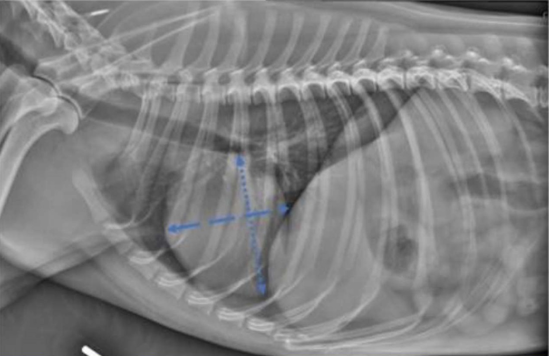
Fig. 1. Principle of vertebral heart scale measurement [1]
The long axis of the heart is measured from the ventral border of the left main bronchus to the most remote ventral point of the apex of the heart. The short axis is measured along a line perpendicular to the long axis, at the level of the caudal vena cava. The sum of the lengths of the two axes is compared to the length of the vertebrae, starting from the cranial edge of T4 (the fourth thoracic vertebra) [2]. This technique has been proposed as one of the methods for assessing cardiomegaly in dogs and cats.
In 2001, H. Nakayama et al. conducted a study to determine the extent the echocardiography and electrocardiography results correlate with VHS. The experiment involved dogs with cardiomegaly of different severity. They were monitored using chest radiography, echocardiography, and electrocardiography in dynamics. Then, echocardiographic and electrocardiographic parameters were compared to VHS. The results of the study showed that the Left Atrial to Aorta ratio, left ventricular end-diastolic volume and end-systolic volume, P-wave duration and QRS interval duration correlated consistently with VHS [3].
However, the use of the vertebral heart scale (VHS) measurement method has its limitations. Firstly, although it enables quite objective determination of the presence of cardiomegaly in animals, it does not provide an opportunity to assess the entire area of the heart: VHS is a one-dimensional method and has functional limitation, as it uses only two linear measurements to determine the heart size, and not the entire heart circumference. Secondly, significant limitations are related to the interpretation of images, which is subjective in nature, since it largely depends on the education and experience of a radiologist (common causes of misinterpretation are perception errors, omissions resulting from the lack of attention, cognitive distortions, fatigue, distraction). Certain steps are currently taken to integrate the artificial intelligence (AI) technologies into X-ray diagnostics: this might provide a number of advantages, including the possibility of computerized interpreting complex images and calculating certain parameter. An experiment on measuring vertebral heart scale in dogs and cats by means of AI was held in 2021[1].
The aim of the present review is to summarise and analyse data available in the literature on the use of vertebral heart scale measurement during chest radiography in cats and dogs, to assess the relevance of this technique and prospects of development.
Materials and Methods. The search for scientific articles for the review was carried out in the PubMed (www.pubmed.ncbi.nlm.nih.gov/) and eLIBRARY.RU (www.elibrary.ru/defaultx.asp) databases. Only works in Russian and English published between January 1, 1995 and December 31, 2024 were included in the search. In PubMed, the search was conducted using the keywords: ((radiomics or X-ray) and (Vertebral Heart Scale or VHS) and (radiographs or diagnostic imaging) and (cats) and (dogs)). The search for scientific publications in eLIBRARY.RU was carried out using the option of “advanced search” was used by entering the following combination of words into the dialog box: ((X-ray or X-ray diagnostics) and (Vertebral Heart Scale or VHS) and (cats and dogs or small domestic animals)). In the “Where to search?” box, the search was specified by the criteria: “in the title”, “in the abstract”, “in keywords”. “Publication type” was indicated as “journal articles”. Additional search criteria were the parameters “search taking into account morphology” and “search in publications that have full text on eLIBRARY.RU”.
A scientific paper was included in the review based on the following criteria: 1) the article had to contain the information on the use of vertebral heart scale method of measurement in dogs or cats, 2) the article had to have a full-text version. Non-conformity of a literature source with the criteria for inclusion in the review was the reason for its exclusion. The results were presented in the form of a PRISMA flow chart and a table.
Research Results. In total, 63 sources were found as a result of the database search. On the whole, 13 duplicating sources were excluded, and 50 articles were selected for further analysis. After screening the titles and abstracts, 5 sources were removed. Potentially eligible full texts (n = 45) were uploaded to cloud storage and reviewed by all the experts to ensure their relevance. After this step, 14 more publications were excluded. The final number of articles selected for inclusion in the review was 31. A summary of the screening process is shown in the PRISMA flow chart (Figure 2).
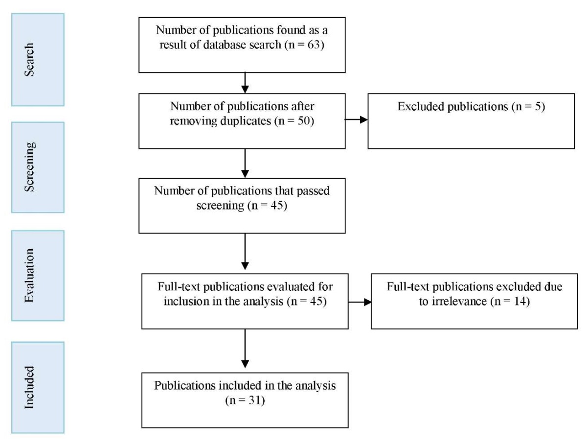
Fig. 2. PRISMA flow chart with the results of publication selection process
The majority of scientific publications (28 articles) referred to vertebral heart scale measurement in dogs and cats in the normal and pathological condition; 2 articles investigated the requirements to the technique of acquiring X-ray images intended for measuring VHS value; 1 article studied the use of artificial intelligence technology for measuring vertebral heart scale in dogs and cats.
The use of vertebral heart scale method of measurement for assessing the size of the cardiac silhouette during radiodiagnostics of dogs was first mentioned in 1995 [2]. J.W. Buchanan and J. Bücheler developed this method based on the fact that there was a good correlation between the size of the heart and the length of vertebral bodies regardless of the shape of canine thorax. 100 clinically healthy dogs at the age over one year (regardless of sex) were selected for the study. On the obtained radiographs of the cardiac silhouette, the sizes of the long and short axes were determined using a beam caliper, summed up and compared with the length of the vertebrae, starting from T4: the ratio was 9.7 ± 0.5 vertebra. Differences between the values in dogs with a wide or deep chest, males and females, as well as between the right or left lateral views were insignificant and were not taken into account. Afterwards, the obtained VHS value was for a long time considered the norm for the dogs of various breeds [2].
Later, in 2000, a similar study involved 100 clinically healthy cats. In the lateral X-rays, the average heart size relative to the vertebrae was 7.5 ± 0.3 vertebrae. The data obtained made it possible to conclude that the method for determining the size of the heart relative to the vertebrae is easy to use. It enables objective assessment of the heart size, thus, can be helpful in determining the presence of cardiomegaly as well as in comparing the sizes of the cardiac silhouette on a series of consecutive radiographs. In addition, this study showed that the VHS method of measurement is effective not only in dogs, but also in cats [4].
The earliest experiments of VHS measurement involved only healthy animals. Therefore, to evaluate the capacity of vertebral heart scale measurement for the description of chest X-rays in animals with cardiac pathology, another study was conducted in 2000: chest X-rays of 50 dogs with established cardiac pathology, 26 Х-rays of dogs with respiratory pathology, and 50 Х-rays of dogs with no clinical signs of cardiovascular or respiratory diseases were mixed and examined by three veterinarians. In the first examination of the X-rays, VHS was not measured, and the diagnosis was made based on other radiographic features. The accuracy of cardiac disease diagnosis by each expert based on the subjective evaluation of radiographs without VHS measurement was moderately accurate (ranging from 76% to 83% of correct diagnoses). Afterwards, the experts re-examined the radiographs and measured VHS on both lateral and dorsoventral or ventrodorsal radiographs. It is worth noting that measuring vertebral heart scale on the examined radiographs did not almost influence the decision about correctness of the previously made diagnosis. However, all veterinarians got a higher mean VHS value in dogs with heart pathology than in dogs with other pathologies or without them. Based on the results of the experiment, it was concluded that VHS value more than 10.7 on the lateral X-rays can be considered a fairly consistent sign of heart pathology [5].
In 2001, the vertebral heart scale was measured in 11 puppies without signs of pathology. It was found that the standards for determining the indicators of enlarged heart in puppies and adult dogs were similar [6].
Later on, due to the large variability of breed characteristics in dogs, the studies on measuring the vertebral heart scale were carried out individually for each breed. In 2005, the VHS was measured for 44 dogs of Whippet breed having no heart and lung diseases. The mean vertebral heart scale value for this breed upon evaluation of the right lateral X-rays was 11.3 ± 0.5 vertebrae [7].
In 2007, the measurement was carried out in Greyhounds. It aimed to compare VHS in normal Greyhounds to that in Rottweilers and a group of dogs of other breeds. As a result, it was found that VHS was significantly higher in Greyhounds compared to Rottweilers and other breeds. For Greyhounds, the mean VHS on lateral radiographs was 10.5 ± 0.1, for Rottweilers — 9.8 ± 0.1, and for mixed breed dogs — 10.1 ± 0.2. This study also confirmed that relative cardiomegaly, subsequently detected at necropsy and ECG in Greyhounds, could be easily detected by plain radiography [8].
In 2007, radiographs of 50 adult stray cats were examined. All cats were short-haired, non-obese, and considered healthy based on clinical examination and electrocardiography. Long and short axes of heart were measured in millimetres. Length of vertebrae was measured caudally from T4. The sum of the long and short axes of the heart expressed as VHS was 7.3 ± 0.49 vertebrae in the right lateral view and 7.3 ± 0.55 vertebrae in the left lateral view. Differences between the right and left lateral views were minor [9].
In 2008, 19 Beagle dogs were examined using echocardiography, electrocardiography, noninvasive blood pressure measurement, complete blood count, and serum chemistry profile. Then, all studied dogs underwent the right and left lateral chest radiography, and vertebral heart scale measurement. The VHS in Beagles was 10.3 ± 0.4 vertebrae, which differed from the mean VHS value of 9.7 ± 0.5 as determined in JW Buchanan’s original study for dogs of different breeds [2]. The data obtained had once again confirmed the fact that the results should be interpreted taking into account breed-specific differences, especially for those values that were significantly different from the reference range [10].
In 2009, the efficiency of VHS measurement was evaluated in dogs with cough and established degenerative mitral valve disease. Chest X-rays of 90 dogs with coughing in history and echocardiographic evidence of mitral valve disease were examined by two independent specialists. They were first asked to determine the cause of cough (cardiogenic, non-cardiogenic, or mixed) and then to measure VHS. Dogs with non-cardiogenic cough had significantly lower VHS (mean value 11.0 ± 0.9) compared to the dogs with cardiogenic or mixed cough (12.8 ± 1 and 12.9 ± 0.9, respectively). Analysis of the results showed that VHS value less than or equal to 11.4 is a precise enough indication to exclude cough of cardiac origin in dogs with mitral valve disease. The results of the study suggest that VHS measurement may be an additional tool for differentiating the cause of coughing in dogs with cardiac diseases [11].
In 2013, a study was published describing the measurement of VHS in dogs of the following breeds: Pug, Pomeranian, Yorkshire Terrier, Dachshund, Bulldog, Shih Tzu, Lhasa Apso, and Boston Terrier. The study included healthy animals with no subjective radiographic signs of cardiomegaly, without murmurs, or abnormal heart rhythms during clinical examination. It was found that in groups of dogs that included Pugs, Pomeranians, Bulldogs, and Boston Terriers, mean VHS was 10.7 ± 0.5 [12].
The aim of another experiment in the same 2013 was to evaluate the efficiency of VHS method in differentiating congestive heart failure from other causes of dyspnea in cats. To be included in the study, cats were examined using echocardiography and chest radiography within 12 hours after admission to the clinic. The study revealed that VHS > 9.3 vertebrae was a differentiating feature for detection of cardiac diseases. Measurements, which resulted in the range of 8.0 to 9.3 suggested that the cause of dyspnea might be equivocal (i.e. secondary to congestive heart failure or respiratory disease), in this case echocardiography would be the most useful method in providing additional diagnostic information. The results of the study had revealed that VHS method of measurement may be a useful tool facilitating differentiation of cardiogenic from non-cardiogenic causes of respiratory failure in cats in the emergency situation when echocardiography is unavailable or not warranted in an unstable patient [13].
In 2014, echocardiographic and radiographic examination of thorax was carried out in 150 cats aiming to evaluate the diagnostic accuracy of VHS and other related radiographic indices upon detecting cardiomegaly caused by various feline cardiac diseases. Eighty-three cats had various cardiac diseases, and 22 cats had no cardiovascular anomalies. The measurement yielded the following values: VHS in the lateral view in healthy cats was 7.56 ± 0.54; in cats with left-sided heart diseases — 8.62 ± 1.04; in cats with left-sided heart diseases but without left atrial enlargement or with moderate left atrial enlargement — 8.29 ± 0.83; in cats with left-sided heart diseases and with moderate to severe left atrial enlargement — 8.95 ± 1.13 [14].
In 2016, a study was conducted to establish reference values for VHS in Indian Spitz, Labrador Retrievers, and outbred dogs. In total, 60 animals (20 Indian Spitz, 20 Labrador Retrievers, and 20 outbred dogs) with no radiographic or clinical signs of cardiovascular or pulmonary diseases were included in the study. As a result of the study, the following VHS values in the right lateral view were established: 10.21 ± 0.13 — for Spitz breed, 10.39 ± 0.19 — for Labrador Retrievers, and 9.8 ± 0.21 — for outbred dogs [15].
In 2017, an experiment aimed at measuring the vertebral heart scale in Dachshund dog breed and comparing the results with the established reference range of 9.7 ± 0.5 was conducted. It involved 51 dogs of Dachshund breed, which underwent radiography and echocardiography. As a result of the study, it was found that mean VHS in the right lateral X-ray was 10.3 vertebrae (the range 9.25–11.55). Whereas VHS in females was higher than in males: 10.8 (9.50–11.55) versus 9.99 (9.25–10.8). The results revealed that in healthy Dachshunds, the mean VHS value exceeded the published standard range for dogs [16].
In 2019, vertebral heart scale was measured in 75 dogs with preclinical stage of atrioventricular valve endocardiosis. The study revealed that, with this pathology, increase of vertebral heart scale value confirmed the presence of cardiomegaly, but did not specify the parts of the heart with pathological changes [17].
In another study of 2019 VHS was measured in 61 dogs of Norwich Terrier breed without clinical signs of cardiovascular disease. It was found that VHS value in dogs of this breed was 10.6 ± 0.6, which is higher than the reference value for dogs (9.7 ± 0.5) [18].
Also in 2019, the VHS was measured in the Australian Cattle dog breed. The study involved 20 dogs having no heart or lung diseases. The mean VHS value in the Australian Cattle Dog turned to be also higher (10.5 ± 0.4) compared to the mean VHS originally indicated for 100 healthy dogs of different breeds (9.7 ± 0.5) [19].
In the study held in 2021, mean reference VHS value was established in Persian cats equalling to 8.16 vertebrae (higher than the value of 7.5 reported in earlier studies for other cat breeds). It was found that gender did not affect these values [20].
In the same year, 2021, VHS was measured in Maltese dog breed. It involved 81 clinically healthy dogs that had undergone a complete cardiac examination. The mean VHS was 9.53 ± 0.46 vertebrae [21].
At the end of 2021, VHS was measured in Brittany Spaniel dog breed. The study included 28 healthy dogs and 15 dogs with myxomatous mitral valve disease. Measurements were taken by examining the right lateral X-rays. The mean VHS ± SD (standard deviation) was 10.6 ± 0.2 in healthy animals and 11.9 ± 1.1 in animals with identified pathology [22].
Another paper of 2021 encompassed VHS measurement in 30 dogs of Chihuahua breed. The aim of the study was to assess VHS value in healthy adult dogs, since this breed is predisposed to both congenital and acquired heart diseases. The VHS value in the sample population was 10.0 ± 0.6 [23].
In 2022, VHS was established for Pembroke Welsh Corgi dog breed and the influence of the thoracic vertebrae features on this measurement was assessed. As a result, it was possible to measure the vertebral heart scale in these animals, which equalled to 9.36 ± 0.27 (below the reference value). The authors supposed that it might be due to the slight difference of the features of thoracic vertebrae in Corgi from other breeds [24].
In 2022, a study involving dogs of Cavalier King Charles Spaniel breed was conducted. This breed is predisposed to developing myxomatous mitral valve disease, and radiographs are often used to detect signs of left-sided cardiomegaly secondary to this disease. The study was conducted in 30 clinically healthy adult dogs (22 females and 8 males), aged 1 to 6 years. Inclusion criteria were: absence of pathologies during physical examination, normal echocardiography, and chest radiographs without signs of anomaly. The mean VHS value in the sample was 10.08 ± 0.56. This value exceeded the previously published general reference value for dogs equal to 9.7 ± 0.5 vertebrae [25].
In the earlier experiments of 2021, VHS was measured for healthy dogs of Chihuahua breed. The aim of the study conducted in 2022 was to evaluate the influence of cardiac enlargement on VHS in Chihuahuas with myxomatous mitral valve disease. The experiment included 10 clinically normal Chihuahuas and 97 Chihuahuas with the disease. The sick dogs were divided into 3 groups depending on the severity of the disease: mild, moderate, severe. Mean VHS in healthy dogs was 9.66 ± 0.36; in those with mild myxomatous mitral valve disease — 10.13 ± 0.64; with moderate — 10.87 ± 0.71; with severe — 11.71 ± 0.78. Conclusion: VHS in dogs of this breed increased depending on the cardiac enlargement caused by myxomatous mitral valve disease [26].
In 2023, a chest X-ray examination was performed in 10 dogs diagnosed with dilated cardiomyopathy (DCM). Measurement of vertebral heart scale showed that, on average, its value increased by 0.5–1.0, which was a consistent indication of the presence of cardiomegaly confirmed by other methods of visual diagnostics as well [27].
In the same 2023, by measuring VHS in 29 clinically healthy dogs it was possible to establish its value for American Staffordshire Terrier dog breed. Each dog underwent ECG, chest X-ray and echocardiography. All dogs had no cardiac or pulmonary pathologies. The mean VHS value for this breed was 10.9 ± 0.6. This value did not differ significantly between males and females, and did not have consistent correlation with age or body weight of an animal [28].
In late 2023, the expediency of measuring VHS for predicting left-sided congestive heart failure in dogs with respiratory signs was determined. The study included 114 dogs with respiratory signs and radiographic evidences of lung disorders. Animals underwent chest echocardiography and radiography. The diagnosis of left-sided congestive heart failure was confirmed based on the presence of respiratory signs, left ventricular hypertrophy, and cardiogenic pulmonary edema. In half of the animals participating in the study this pathology was established, and in the other half — it was not. According to the results of VHS measurement in animals without pathology, its mean value was 11.1 (10.1–11.8), and in dogs diagnosed with this pathology, it was 12.9 (11.9–14.1). These results may help to exclude cardiogenic pathology in dogs with respiratory signs [29].
VHS values for different dog breeds are presented in Table 1, indicating the year of the study.
Table 1
Vertebral heart scale values for different dog breeds
|
Dog breed |
Year of the study |
Established VHS Value |
|
Whippet |
2005 |
11,3 ± 0,5 |
|
Greyhound |
2007 |
10,5 ± 0,1 |
|
Rottweiler |
2007 |
9,8 ± 0,1 |
|
Beagle |
2008 |
10,3 ± 0,4 |
|
2015 |
10,59 ± 0,49 (right) and 10,35 ± 0,50 (left) during the inhalation phase. 10,11 ± 0,37 (right) and 9,92 ± 0,50 (left) during the inhalation phase |
|
|
Pug, Pomeranian, Shih Tzu, Yorkshire Terrier, Lhasa Apso, English Bulldog, Boston Terrier |
2013 |
10,7 ± 0,5 |
|
Indian Spitz |
2016 |
10,21 ± 0,13 |
|
Labrador Retriever |
2016 |
10,39 ± 0,19 |
|
Dachshund |
2017 |
10,3 |
|
Norwich Terrier |
2019 |
10,6 ± 0,6 |
|
Australian Cattle Dog |
2019 |
10,5 ± 0,4 |
|
Maltese |
2021 |
9,53 ± 0,46 |
|
Brittany Spaniel |
2021 |
10,6 ± 0,2 |
|
Chihuahua |
2021 2022 |
10,0 ± 0,6 9,66 ± 0,36 |
|
Pembroke Welsh Corgi |
2022 |
9,36 ± 0,27 |
|
Cavalier King Charles Spaniel |
2022 |
10,08 ± 0,56 |
|
American Staffordshire Terrier |
2023 |
10,9 ± 0,6 |
The review also included literature sources studying the requirements to the technique of acquiring X-ray images and the prospects of using artificial intelligence technology to measure VHS.
In 2008, the paper describing the effect of using the left or right lateral view in dogs for measuring VHS was published. The study was conducted on 63 healthy dogs. VHS was slightly higher in the right lateral decubitus position (9.8 ± 0.6) compared to the left position (9.5 ± 0.8). The sex and size of a dog did not significantly affect VHS values [30].
In 2015 the study was conducted aimed to evaluate the variability of VHS values caused by cardiac and respiratory cycles in Beagle dog breed. The experiment involved 14 dogs that underwent X-ray examination, VHS was measured during the inhalation phase and exhalation phase, and during systole and diastole in the left and right lateral decubitus positions. The mean VHS value was compared within and between cardiac and respiratory phases. The mean VHS for each phase of the respiratory and cardiac cycle was higher on images obtained in the right lateral decubitus position compared to the left lateral decubitus position. The largest differences were observed between VHS in the diastolic phase of inhalation (10.59 ± 0.49 and 10.35 ± 0.50 for the right and left lateral views, respectively) and systolic phase of exhalation (10.11 ± 0.37 and 9.92 ± 0.50 for the right and left lateral views, respectively). The results of the study emphasise the need for the clinicians to be aware of the potential influence of these factors when assessing VHS in dogs [31]
A forward-looking direction for X-ray diagnostics in veterinary medicine is the use of artificial intelligence (AI) technology for measuring vertebral heart scale. In the study of 2021, scientists compared the results of VHS measurement in cats and dogs performed by artificial intelligence and two certified specialists [1]. Thirty canine lateral chest X-rays and thirty feline lateral chest X-rays were used. The images were assessed by each specialist using two different methods for measuring the short axis of heart on canine radiographs: the original method published by Buchanan J.W. and a modified method. Whereas feline radiographs, were assessed using only Buchanan J.W. method. On the whole, VHS values measured by artificial intelligence, a radiologist, and a cardiologist had a high degree of agreement in both dogs and cats (intraclass correlation coefficient (ICC) = 0.998). The ability of the AI trained to find VHS reference values was consistent with the manual measurements carried out by the human specialists for both cats and dogs. The present study demonstrates an important benefit of such a computerized method for general veterinarians as it limits variability in the interpretation of results and provides more comparable VHS values over time [1].
Discussion and Conclusion. A review and analysis of scientific literature on application of vertebral heart scale measurement method in dogs and cats during X-ray diagnostics of thoracic cavity enables to draw the following conclusions:
1. Boissady E, De La Comble A, Zhu X, Abbott J, Adrien-Maxence H. Comparison of a Deep Learning Algorithm vs. Humans for Vertebral Heart Scale Measurements in Cats and Dogs Shows a High Degree of Agreement Among Readers. Frontiers in Veterinary Science. 2021;8:764570. http://doi.org/10.3389/fvets.2021.764570
2. Buchanan JW, Bücheler J. Vertebral Scale System to Measure Canine Heart Size in Radiographs. Journal of the American Veterinary Medical Association. 1995;206(2):194–199. http://doi.org/10.2460/javma.1995.206.02.194
3. Nakayama H, Nakayama T, Hamlin RL. Correlation of Cardiac Enlargement as Assessed by Vertebral Heart Size and Echocardiographic and Electrocardiographic Findings in Dogs with Evolving Cardiomegaly Due to Rapid Ventricular Pacing. Journal of Veterinary Internal Medicine. 2001;15(3):217–221. https://doi.org/10.1892/08916640(2001)015%3C0217:coceaa%3E2.3.co;2
4. Litster AL, Buchanan JW. Vertebral Scale System to Measure Heart Size in Radiographs of Cats. Journal of the American Veterinary Medical Association. 2000;216(2):210–214. http://doi.org/10.2460/javma.2000.216.210
5. Lamb CR, Tyler M, Boswood A, Skelly BJ., Cain M. Assessment of the Value of the Vertebral Heart Scale in the Radiographic Diagnosis of Cardiac Disease in Dogs. Veterinary Record. 2000;146(24):687–690. http://doi.org/10.1136/vr.146.24.687
6. Ghadiri A, Avizeh R, Rasekh A, Yadegari A. Radiographic Measurement of Vertebral Heart Size in Healthy Stray Cats. Journal of Feline Medicine and Surgery. 2008;10(1):61–65. https://doi.org/10.1016/j.jfms.2007.06.015
7. Sleeper M.M, Buchanan JW. Vertebral Scale System to Measure Heart Size in Growing Puppies. Journal of the American Veterinary Medical Association. 2001;219(1):57–59. https://doi.org/10.2460/javma.2001.219.57
8. Bavegems V, Van Caelenberg A, Duchateau L, Sys SU, Van Bree H, De Rick A. Vertebral Heart Size Ranges Specific for Whippets. Veterinary Radiology & Ultrasound. 2005;46(5):400–403. https://doi.org/10.1111/j.17408261.2005.00073.x
9. Marin LM, Brown J, McBrien C, Baumwart R, Samii VF, Couto CG. Vertebral Heart Size in Retired Racing Greyhounds. Veterinary Radiology & Ultrasound. 2007;48(4):332–334. https://doi.org/10.1111/j.1740-8261.2007.00252.x
10. Kraetschmer S, Ludwig K, Meneses F, Nolte I, Simon D. Vertebral Heart Scale in the Beagle Dog. Journal of Small Animal Practice. 2008;49(5):240–243. http://dx.doi.org/10.1111/j.1748-5827.2007.00531.x
11. Guglielmini C, Diana A, Pietra M, Di Tommaso M, Cipone M. Use of the Vertebral Heart Score in Coughing Dogs with Chronic Degenerative Mitral Valve Disease. Journal of Veterinary Medical Science. 2009;71(1):9–13. https://doi.org/10.1292/jvms.71.9
12. Jepsen-Grant K, Pollard RE, Johnson LR. Vertebral Heart Scores in Eight Dog Breeds. Veterinary Radiology & Ultrasound. 2013;54(1):3–8. https://doi.org/10.1111/j.1740-8261.2012.01976.x
13. Sleeper MM, Roland R, Drobatz KJ. Use of the Vertebral Heart Scale for Differentiation of Cardiac and Noncardiac Causes of Respiratory Distress in Cats: 67 Cases (2002–2003). Journal of the American Veterinary Medical Association. 2013;242(3):366–371. http://dx.doi.org/10.2460/javma.242.3.366
14. Guglielmini C, Toaldo MB, Poser H, Menciotti G, Cipone M, Cordella A, Contiero B., Diana A. Diagnostic Accuracy of the Vertebral Heart Score and Other Radiographic Indices in the Detection of Cardiac Enlargement in Cats with Different Cardiac Disorders. Journal of Feline Medicine and Surgery. 2014;16(10):812–825. https://doi.org/10.1177/1098612x14522048
15. Bodh D, Hoque M, Saxena AC, Gugjoo MB, Bist D, Chaudhary JK. Vertebral Scale System to Measure Heart Size in Thoracic Radiographs of Indian Spitz, Labrador Retriever and Mongrel Dogs. Veterinary World. 2016;9(4):371–376. http://doi.org/10.14202/vetworld.2016.371-376
16. Birks R, Fine DM, Leach SB, Clay SE, Eason BD, Britt LG. et al. Breed-Specific Vertebral Heart Scale for the Dachshund. Journal of the American Animal Hospital Association. 2017;53(2):73–79. http://doi.org/10.5326/JAAHA-MS-6474
17. Annikov VV, Annikova LV, Yegunova AV, Mikhalkin AS, Pantyulin AM, Shaykhraziyeva ESh. Dynamics of Changes in Thoracic Radiographs in Dogs with Endocardial AV Valves at the Preclinical Stage during Therapy with an Ace Inhibitor and an Aldosterone Antagonist. In: Proceedings of the XII International Scientific and Practical Conference “Innovative technologies in science and education”. Penza, July 05, 2019. Penza: Nauka i Prosveshchenie (IP Gulyaev G.Yu.) Publ.; 2019. P. 324–329. (In Russ.)
18. Taylor CJ, Simon BT, Stanley BJ, Lai GP, Thieman Mankin KM. Norwich Terriers Possess a Greater Vertebral Heart Scale than the Canine Reference Value. Veterinary Radiology & Ultrasound. 2020;61(1):10–15. https://doi.org/10.1111/vru.12813
19. Luciani MG., Withoeft J.A., Cardoso Pissetti H.M., de Souza L.P., Sombrio M.S., Bach E.C. et al. Vertebral Heart Size in Healthy Australian Cattle Dog. Anatomia, Histologia, Embryologia. 2019;48(3):264–267. https://doi.org/10.1111/ahe.12434
20. Sak D, Pazvant G. Estimation of Vertebral Heart Size and Cardiothoracic Ratio in Persian Cats. Anatomia, Histologia, Embryologia. 2021;50(3):543–549. https://doi.org/10.1111/ahe.12659
21. Baisan RA, Vulpe V. Vertebral Heart Size and Vertebral Left Atrial Size Reference Ranges in Healthy Maltese Dogs. Veterinary Radiology & Ultrasound. 2022;63(1):18–22. https://doi.org/10.1111/vru.13027
22. Kallassy A, Calendrier E, Bouhsina N, Fusellier M. Vertebral Heart Scale for the Brittany Spaniel: Breed-Specific Range and Its Correlation with Heart Disease Assessed by Clinical and Echocardiographic Findings. Veterinary Sciences. 2021;8(12):300. https://doi.org/10.3390/vetsci8120300
23. Puccinelli C, Citi S, Vezzosi T, Garibaldi S, Tognetti R. A Radiographic Study of Breed-Specific Vertebral Heart Score and Vertebral Left Atrial Size in Chihuahuas. Veterinary Radiology & Ultrasound. 2021;62(1):20–26. https://doi.org/10.1111/vru.12919
24. Tangpakornsak T, Saisawart P, Sutthigran S, Jaturunratsamee K, Tachampa K, Thanaboonnipat C, et al. Thoracic Vertebral Length-to-Height Ratio, a Promising Parameter to Predict the Vertebral Heart Score in Normal Welsh Corgi Pembroke Dogs. Veterinary Sciences. 2023;10(2):168. https://doi.org/10.3390/vetsci10020168
25. Bagardi M, Locatelli C, Manfredi M, Bassi J, Spediacci C, Ghilardi S. et al. Breed-Specific Vertebral Heart Score, Vertebral Left Atrial Size, and Radiographic Left Atrial Dimension in Cavalier King Charles Spaniels: Reference Interval Study. Veterinary Radiology & Ultrasound. 2022;63(2):156–163. https://doi.org/10.1111/vru.13036
26. Ito D. Vertebral Heart Size is Associated with Cardiac Enlargement in Chihuahuas with Myxomatous Mitral Valve Disease. Canadian Veterinary Journal. 2022;63(6):627–632.
27. Senchuk IV, Mozar KA, Malginov DE. The Importance of Instrumental Research Methods in the Complex Diagnosis of Dilated Cardiomyopathy in Dogs. Izvestiya sel'skokhozyaistvennoi nauki Tavridy. 2023;(33(196)):115–124. (In Russ.)
28. Szpinda O, Parzeniecka-Jaworska M, Czopowicz M, Jońska I, Bonecka J, Jank M. Cardiological Reference Intervals in Adult American Staffordshire Terrier Dogs. Animals. 2023;13(15):2436. https://doi.org/10.3390/ani13152436
29. Ross ES, Visser LC, Sbardellati N, Potter BM, Ohlendorf A, Scansen BA. Utility of Vertebral Left Atrial Size and Vertebral Heart Size to Aid Detection of Congestive Heart Failure in Dogs with Respiratory Signs. Journal of Veterinary Internal Medicine. 2023;37(6):2021–2029. https://doi.org/10.1111/jvim.16918
30. Greco A, Meomartino L, Raiano V, Fatone G, Brunetti A. Effect of Left vs. Right Recumbency on the Vertebral Heart Score in Normal Dogs. Veterinary Radiology & Ultrasound. 2008;49(5):454–455. https://doi.org/10.1111/j.17408261.2008.00406.x
31. Olive J, Javard R, Specchi S, Bélanger MC, Bélanger C, Beauchamp G, et al. Effect of Cardiac and Respiratory Cycles on Vertebral Heart Score Measured on Fluoroscopic Images of Healthy Dogs. Journal of the American Veterinary Medical Association. 2015;246(10):1091–1097. https://doi.org/10.2460/javma.246.10.1091
Yulia S. Shmarenkova - Senior Lecturer of the Veterinary Medicine and Animal Physiology Department, Faculty of Veterinary Medicine and Animal Science, Kaluga Branch of the Russian State Agrarian University — Moscow Agricultural Academy Named after K.A. Timiryazev.
27, Vishnevsky Str., Kaluga, 248007
Sergey V. Akchurin - Dr. Sci. (Veterinary Medicine), Professor of the Veterinary Medicine Department, Institute of Animal Science and Biology, Russian State Agrarian University — Moscow Agricultural Academy Named after. K.A. Timiryazev.
49, Timiryazevskaya Str., Moscow, 127434
Margarita A. Gabedava - Cand. Sci. (Agricultural Science) Associate Professor of the Veterinary Medicine and Animal Physiology Department, Faculty of Veterinary Medicine and Animal Science, Kaluga Branch of the Russian State Agrarian University —Moscow Agricultural Academy Named after K.A. Timiryazev.
27, Vishnevsky Str., Kaluga, 248007
Tatyana A. Spasskaya - Cand. Sci. (Biology), Associate Professor of the Veterinary Medicine and Animal Physiology Department, Faculty of Veterinary Medicine and Animal Science, Kaluga Branch of the Russian State Agrarian University —Moscow Agricultural Academy Named after K.A. Timiryazev.
27, Vishnevsky Str., Kaluga, 248007
Olga A. Voronkova - Cand. Sci. (Agricultural Science), Associate Professor of the Veterinary Medicine and Animal Physiology Department, Faculty of Veterinary Medicine and Animal Science, Kaluga Branch of the Russian State Agrarian University —Moscow Agricultural Academy Named after K.A. Timiryazev.
27, Vishnevsky Str., Kaluga, 248007
X-ray examination remains an important diagnostic tool in veterinary medicine. The article presents new data on Vertebral Heart Scale measurement that improves assessment of the cardiac silhouette in dogs and cats. The capacity of artificial intelligence technologies in improvement of VHS measurement accuracy was shown. The study confirms the reliability of this method for diagnosis of cardiomegaly and suggests directions for further research.
Shmarenkova Yu.S., Akchurin S.V., Gabedava M.A., Spasskaya T.A., Voronkova O.A. Using the Vertebral Heart Scale Method of Measurement during X-Ray Diagnostics of Thoracic Cavity in Dogs and Cats: A Literature Review. Russian Journal of Veterinary Pathology. 2025;24(2):43-54. https://doi.org/10.23947/2949-4826-2025-24-2-43-54. EDN: GPKBZO
ISSN 2949-4826 (online)
Contact with: Publisher / Editorial Office of the Journal
Publisher: Don State Technical University - DSTU, Rostov-on-Don, Russian Federation - https://donstu.ru/en/
Editor-in-Chief: Alexey М. Ermakov, Dr. Sci. (Biology), Professor, Don State Technical University (Rostov-on-Don, Russian Federation)


