PARASITOLOGY
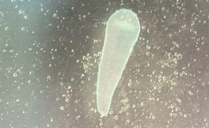
For the first time, the paper presents a study on the infestation of Black Sea-Caspian sprat (Clupeonella cultriventris) with cestodes of the genus Proteocephalus in the Kama Reservoir, and provides the results of conducted histological examination of the intestines of infested fish. It was found that the highest infestation extensity (IE) (50%) was registered in the central area of the reservoir, whereas fish from the lower area was not infested. Infestation with cestodes affected the size and weight of sprat: the weight and length of specimens from the central area were less than of fish from uninfested areas. The histological analysis revealed reversible pathological changes of inflammatory nature in the mucosa and submucosa of the intestines. The discovery of the pre-adult forms of cestodes indicates the need for further research to clarify the parasite-host relationships. The data obtained are important for understanding the role of the alien sprat in spreading parasites and for assessing its status quo in the Kama Reservoir ecosystem.
Introduction. The Black Sea-Caspian sprat (Clupeonella cultriventris) is an alien species adapted to the Kama Reservoir conditions, capable of affecting the trophic relationships in this ecosystem. However, the composition of helminth fauna in this ecosystem and the pathological changes induced by the certain pathogens, such as cestodes of the genus Proteocephalus, are currently poorly investigated. The aim of the study is to investigate the infestation of C. cultriventris with cestodes of the genus Proteocephalus in the Kama Reservoir and to describe the histological changes in the fish intestines caused by this infestation.
Materials and Methods. In the autumn-winter period of 2024, the C. cultriventris (n=83) were taken from the several parts of the Kama Reservoir. A partial biological analysis, partial helminthological dissection and histological examination of the intestines infested with cestodes were carried out. Parasitological examination was performed at the Department of Infectious Diseases of Perm State Agro-Technological University. Preparation of histological specimens was performed in the histopathology laboratory of Perm Regional Children’s Clinical Hospital. The prepared histological sections were scanned using the Vision Assist automated system and Vision microscopy automation software. Mathematical processing of the obtained data was performed in Microsoft Excel.
Results. The highest infestation extensity (IE 50%) of Proteocephalus sp. (juv) corresponding to infestation intensity (II) range 1–2 was observed in sprat form the middle part of the Kama Reservoir; in sprat from the upper part, it was several times less (IE 13% corresponding to II range 1–3); sprat from the lower part was not infested at all. The size and weight parameters in sprat from the upper and lower parts of the Reservoir were significantly higher than that in the fish from the middle part — approximately 2 times exceedance in weight and 1.2 times — in length. Histological examination of sprat intestines revealed changes in mucosa and submucosa layers; the epithelium was in a state of desquamation, the stromal state of the villi was loose and edematous.
Discussion and Conclusions. Data on infestation of C. cultriventris with the cestodes of the genus Proteocephalus sp. in the Kama Reservoir are presented for the first time. It has been revealed that cestode infestation affects the biological parameters of fish and causes minor inflammatory pathological changes in the intestines. Detection of the pre-mature cestodes in the non-specific hosts is not enough factor for their identification, therefore, further research is needed to clarify the host-parasite relationships of C. cultriventris and Proteocephalus sp.
ANIMAL PATHOLOGY, MORPHOLOGY, PHYSIOLOGY, PHARMACOLOGY AND TOXICOLOGY
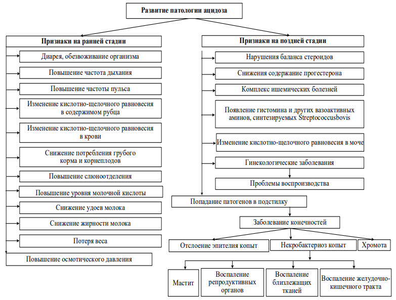
The study presents the first stage of developing an intelligent system for diagnosing rumen acidosis in cows based on fuzzy set theory. The problem was formalized, which included justification of the set of key diagnostic parameters: pH of rumen contents, fat content in milk, pulse rate, and respiratory rate. A hierarchical-type structure of the intelligent system with intermediate variables was developed (“pH–Fat content” and “Respiration–Pulse”), ensuring its logics and transparency. All input, intermediate, and output variables were represented in linguistic terms as specified membership functions. The resulting formalized models will form the basis for the computer implementation of the intelligent system at the second stage of the study, which will enable automation and improve the accuracy of early rumen acidosis diagnostics.
Introduction. Improvement of the diagnostic methods for detecting bovine ruminal acidosis is a relevant problem due to the prevalence of this disease resulting in cattle murrain. The most future-oriented methods of early diagnostics of this pathology are prophylaxis campaigns or individual examinations of animals using the online services in the frame of veterinary telemedicine technologies. However, in Russia, the efficient use of these technologies is hindered by the absence of well-developed methods of assessing the pathology based on the mathematical tools integrated into the advanced intelligent systems. In our study, we attempted to create an intelligent system for diagnosing rumen acidosis in cows. The first stage of the research consisted of formalization of tasks based on the concept of fuzzy logic.
Materials and Methods. The study was conducted at the Ural State Agrarian University and the Northern Trans-Ural State Agricultural University from 2022 to 2025. For diagnosing rumen acidosis, a minimum list of diagnostic parameters was determined as input variables for the fuzzy system (based on the analysis of data obtained from the specialized reference books and scientific publications). Due to the possible difficulty of combining several parameters exceeding reference values, a method for representing uncertainties — fuzzy set theory — was used to correctly solve the problem in the given settings. A hierarchical structure was chosen to build the intelligent system, which makes it strictly logical and transparent for analysis. Scilab open source software with embedded text editor Scinotes were used to develop the application.
Results. A set of necessary diagnostic parameters including pH of rumen contents, fat content in milk, pulse rate, and respiratory rate was justified. A substantive formulation and formalization of the problem of fuzzy inference for assessing rumen acidosis pathology in cows was completed. The structure of the hierarchical-type intelligent system was developed. All variables, including intermediate ones, were formalized within the fuzzy model of the intelligent diagnostic system.
Discussion and Conclusion. In the first part of the study, we have established the formal models of all variables (input, output, and intermediate) for solving the task of diagnosing rumen acidosis in cows. In case of emergence of new parameters directly related to acidosis, they can also be integrated into the intelligent system. Based on the formalized data obtained at this stage of the research, in the second part of the study, it is planned to apply computer technologies to create the intelligent system for assessing rumen acidosis pathology in cows.
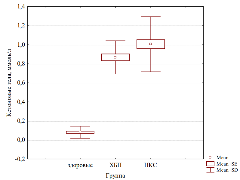
Within the study, a comprehensive analysis of serum biochemical parameters in cats with nephrocardial syndrome (NCS) was conducted compared to the animals with chronic kidney disease (CKD) without cardiac complications and healthy cats. NCS was found to be characterized by pronounced systemic inflammation, manifested in a significant increase of C-reactive protein levels and dysproteinemia: hypoalbuminemia and hyperglobulinemia, and a sharp decrease of the albumin to globulin ratio. Azotemia parameters (urea, creatinine, SDMA) were significantly elevated in cats with both CKD and NCS, however did not differ between these groups, indicating the absence of an additional effect of the cardiac component on the severity of azotemia. Increased ketogenesis, likely associated with uremic intoxication and catabolic processes, was also detected in sick animals. These findings emphasise the importance of integrative assessment of inflammatory and metabolic markers to improve the diagnostics and develop therapeutic strategies for treating nephrocardial syndrome in cats.
Introduction. Nephrocardial syndrome (NCS) is a complex of interrelated disorders, which includes chronic kidney disease (CKD) resulting in development of secondary myocardial dysfunction and structural cardiac remodeling. CKD is one of the most common pathologies in older cats. Although cases of nephrocardial syndrome in cats have been described in the literature, its precise pathophysiological mechanisms are still poorly understood. Particularly insufficient are the scientific data regarding the role of systemic inflammation and protein and energy metabolism disorders related to NCS pathogenesis and progression. The aim of the study is to investigate the biochemical parameters of blood serum in cats with nephrocardial syndrome for identifying the specific patterns of disorders and developing the improved diagnostic and therapeutic techniques.
Materials and Methods. A retrospective analysis of 82 medical records of cats admitted to the Moscow veterinary clinics in the period of 2021 — 2025 was conducted. Two experimental groups of animals were formed (37 cats with NCS; 23 cats with CKD without cardiac complications) and one control group (22 healthy cats). Research methods included serum biochemistry (total protein, albumin/globulin fractions, C-reactive protein, azotemia and ketonemia markers), as well as instrumental methods (echocardiography, ECG).
Results. In cats with NCS, pronounced dysproteinemia was revealed: hypoalbuminemia (28.1±0.5 g/l versus 34.2±0.6 g/l in the control group), hyperglobulinemia (42.6±1.4 g/l), a significant decrease in the albumin to globulin ratio (0.69±0.03 versus 1.02±0.07 in the control group). The level of C-reactive protein in animals with NCS exceeded the values of both healthy cats (164.1±5.6 g/l versus 82.9±1.4 g/l) and the cats from the group with CKD without cardiac complications (128.5±3.2 g/l), which confirms the presence of systemic inflammation. Azotemia parameters (urea, creatinine, SDMA) did not significantly differ in groups with CKD and NCS, but were higher, compared to the control group.
Discussion and Conclusion. The increased ketogenesis was detected in cats with both pathologies, which is likely due to uremic intoxication and catabolic processes. NCS in cats is characterized by pronounced systemic inflammation, impaired protein metabolism, and metabolic shifts. The obtained results emphasize the importance of integrating inflammatory and metabolic markers interpretation into the diagnostics of NCS, as well as the need for further research to establish the cause-and-effect relationships within the nephrocardial continuum.
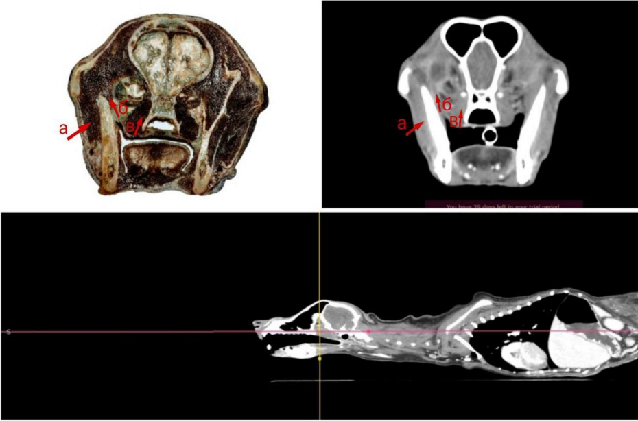
Within the study, a comparative analysis of computed tomography (CT) scans of the masticatory muscles in dogs against the macroscopic anatomy sample sections was conducted to evaluate the accuracy of the technique. CT scans were found to accurately depict the anatomical topography of the masseter and temporalis muscles. Minor discrepancies in the silhouettes of the pterygoid muscles were due to the technical features of freezing and cutting, rather than imaging errors. CT scanning proved its capacity to enable detailed assessment of tissue structure, fiber direction, and tissue density, which is not possible with traditional radiography. High resolution provided by this method, ensures early detection of the inflammatory, traumatic, and neoplastic changes, and facilitates planning the biopsies and surgical interventions. The obtained results confirm that CT scanning is a future-oriented and accurate tool for diagnosing masticatory muscle pathologies in dogs, although further research on a larger sample of clinically confirmed cases is needed to refine the diagnostic criteria.
Introduction. The problem of diagnosing the masticatory muscle pathologies in dogs remains relevant both for veterinary science and practice, as masticatory muscle diseases can lead to the loss of appetite and, as a result, to deterioration of animal overall health. Modern imaging techniques, such as computed tomography (CT) scanning, are still rarely used in veterinary medicine for diagnosing the pathologies. The aim of the present study is to perform CT scanning of the masticatory muscles in dogs and evaluate the diagnostic capacities of this technique by comparing the CT scans of these muscles to macroscopic anatomy sections thereof.
Materials and Methods. The study was conducted at the premises of VitaVet Veterinary Clinic and in the anatomical theatre of Don State Technical University from November 2023 to March 2024. The objects of the study were dogs (n=3). Three-phase CT scans were taken: arterial, venous, and delayed. After CT scanning and euthanasia, the animal cadavers were frozen at the temperature of –20°C for one week. The muscle sections for macroscopic anatomy analysis were obtained using a circular saw table. Alinement of CT images with the actual anatomical structure was established by comparing the macroscopic anatomy sections against the axial and sagittal CT projections of these structures.
Results. The anatomical site and topographic relationships of the masticatory muscles with the associated tissues in canine cadavers were described using computed tomography scanning and macroscopic anatomy analysis of muscle sections. By comparing CT images to the macroscopic anatomy sections, a high degree of matching between them was revealed: the masseter muscle and temporalis muscle silhouettes on CT scans completely matched with their topography in frozen sections. Minor differences in tissue thickness and density were observed in the pterygoid muscles, which could be explained by the features of soft tissue cutting and shrinkage during freezing.
Discussion and Conclusion. Computed tomography scanning is a preferred imaging technique for the masticatory muscles in the head of an animal, as it enables precise determination of the location, quantity, size, and intensity of morphological changes in the muscles reflecting their condition and degree of involvement in the pathological process. However, several constraints should be borne in mind when considering the obtained results: the study was conducted in a small number of animals; the pathologies were not clinically confirmed in the cadavers used for the study, etc. The prospects of future research lie in expansion of the size of a sample to include the animals of different breeds and ages, as well as in incorporation of the clinical cases of myositis, tumours and post-traumatic changes in the masticatory muscles. This will improve the accuracy of diagnostics and efficacy of therapy.
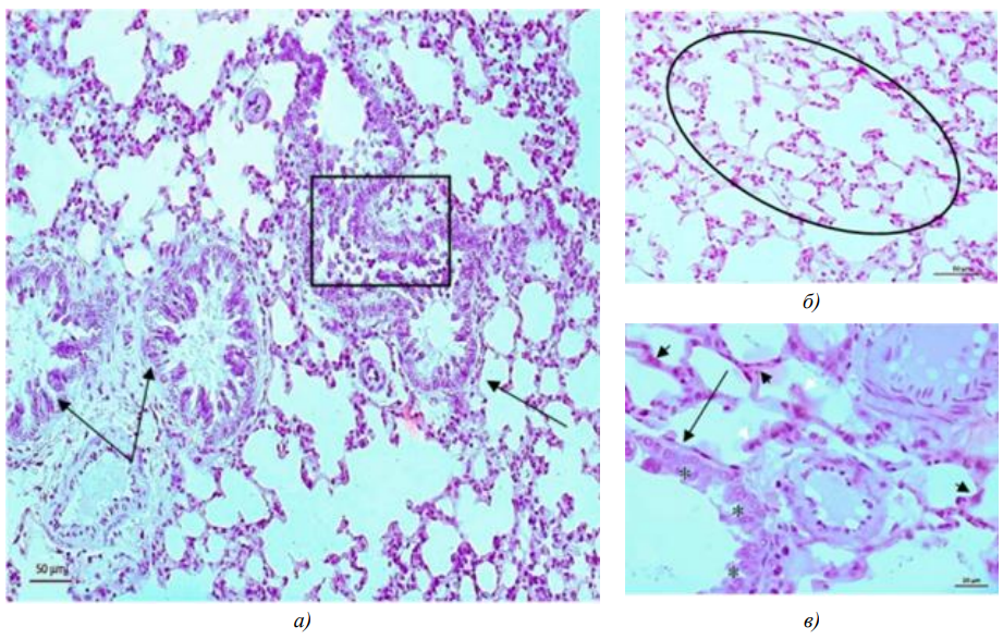
For the first time the morphofunctional changes in the lungs, kidneys, and liver of Djungarian hamsters exposed to long-term (80 days) inhalation of e-cigarette aerosol containing nicotine (3 mg/ml) and flavouring agents were studied. The results demonstrate the pathogenic effects of the multicomponent aerosol, which causes the reversible damage to the bronchial and alveolar epithelium, the disorders of microcirculation and hemodynamics, as well as the signs of functional overload of the organs of detoxification. The revealed changes are similar to those observed in acute respiratory distress syndrome in the lungs and indicate impaired renal filtration-reabsorption function and activation of hepatocyte apoptosis. The study provides the new experimental data on the harm caused by the long-term use of the electronic nicotine delivery systems and emphasises the need for further research to assess their long-term health risks, particularly in the context of their growing popularity.
Introduction. The liquids of electronic nicotine delivery systems (ENDS), which have the multi-component composition, represent a major environmental hazard, as they contain extremely toxic substances. Although the popularity of this form of smoking grows, the data on the biological effects of long-term exposure of animals and humans to e-cigarette and vape components are insufficient. The aim of the present study is to investigate the morphofunctional state of the lungs and main organs of detoxification in Djungarian hamsters under long-term exposure to ENDS aerosol.
Materials and Methods. The research was conducted in 2021 in the colony of laboratory Djungarian hamsters kept at the Institute of Systematics and Ecology of Animals SA RAS. Thirty-six hamsters were divided into two groups. During 80 days, animals in the experimental group (10 females and 10 males) were exposed to vapour from heating the X-3 Yoghurt Pear liquid. The procedure was performed twice a day for 10 minutes, with a 2-hour interval. Hamsters in the control group (8 males and 8 females) were placed in exposure chambers but were not exposed to vapour. At the end of the 80-day experiment, the animals were decapitated, and organs were collected for preparing thin histological sections. The specimens were stained with Boehmer’s hematoxylin and eosin. Collagen distribution was determined using the Mallory method. Histological specimens of organs were examined under transmission light microscope
Results. In experimental animals exposed to toxic vapour, a homogeneous black substance was detected in bronchial epithelial cells and in the alveolar interstitium. In the lung parenchyma, signs of developing interstitial pneumonia, atelectasis, emphysema, and obstructive bronchitis were detected, indicating impaired ventilation-perfusion relationships in the lung tissue and gas exchange disorder. In the kidneys, the homogeneous substance was localized in the lumen of renal tubules. Light optical microscopy revealed the main signs of lethal damage in renal epithelial cells. Strongly expressed dilation of the renal glomerular capillaries, coupled with a nearly 2-fold reduction in the surface area of renal corpuscles compared to control samples, indicates impaired hemodynamics and disrupted renal reabsorption-filtration function. Regarding the liver, a high level of localization of evolutionarily adapted to detoxification dark hepatocytes dying by apoptosis in the centrilobular region of the lobule, indicates the involvement of toxic substances in this process, which enter the liver with the blood.
Discussion and Conclusion. The experiment demonstrated the general pathogenic effects of ENDS aerosol on the lungs and organs of detoxification in animals after long-term exposure to it. Such studies are necessary in the context of the observed growth of consumption of the nicotine delivery systems at the global market.
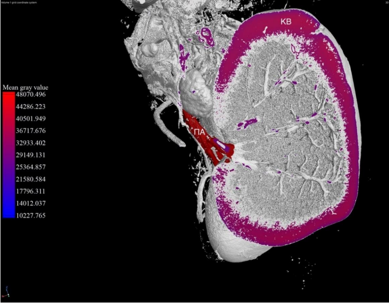
Using computed X-ray microtomography (micro-CT), the study provided the first comprehensive assessment of the changes in kidney tissue density of laboratory rats with induced hyperlipidemia. The method enabled making the high-quality three-dimensional images with a voxel resolution of 20 μm and carrying out quantitative assessment of the dynamics of changes in the cortex, medulla, and vasculature. It was found that the greatest density increase in all these structures occurred after 3 months of a hyperlipidemic diet (peak fat infiltration), while by the end of 5 months, the values approached the control levels, which may indicate the development of fibrotic changes. The results demonstrate the efficiency of micro-CT with regard to non-destructive imaging, accurate quantitative assessment of structural changes, and dynamic monitoring the pathological processes in kidneys during metabolic disorders. The study expands the potential of using micro-CT in veterinary medicine and experimental morphology for the early diagnostics and study of the pathogenesis of internal organ diseases.
Introduction. Computed X-ray microtomography (micro-CT) enables getting the three-dimensional images of microscopic structures without damaging them, which makes this method widely implemented in biomedicine. Regarding the application of micro-CT in veterinary medicine, its potential in assessing morphological changes in internal organs of animals with pathologies has not yet been used at full scale. For example, there are no enough data on the capacity of microtomography in studying pathological processes in kidneys of animals. The aim of the present study is to assess the pathological changes in the density and morphology of kidney tissue in laboratory rats with hyperlipidemia using micro-CT.
Materials and Methods. The experiment was conducted at DSTU from 2021 to 2024 and involved 40 male Wistar rats divided into 5 groups: 4 experimental and 1 control. During the experiment, the experimental groups were fed a hyperlipidemic diet; the control group received only standard feed-stuff. Kidneys were taken from decapitated animals on 30th, 120th, 150th, and 180th day, and samples were prepared for scanning with a Zeiss Xradia Versa 520 micro-CT scanner at 80 kV and voxel size of 20 μm. The efficiency of micro-CT was assessed by the quality of 3D reconstruction and detected changes in kidney tissue density and morphology at different stages of hyperlipidemia.
Results. Microtomograms of kidneys of rats in the experiment allowed for detailed visualization of the organ’s morphology, including renal cortex and medulla, as well as vasculature. Quantitative data on changes in tissue density were obtained, and differences in kidney structure were distinguished between the normal (in control group) and pathological conditions (in experimental groups with various degrees of hyperlipidemia).
Discussion and Conclusion. Micro-CT method has demonstrated high accuracy and informative value in analysing kidney tissue condition in rats and proved its efficiency in early diagnostics of pathological changes in these internal organs, as well as in dynamic monitoring of disease. Among the constraints of this method, the following aspects can be noted: the high cost of equipment, low sensitivity to soft tissues in the absence of contrast-enhancement, and the need for specialized skills to interpret the images.
In the study, the efficiency of modeling the oxidative stress in rats using three prooxidant factors: noise (95–105 dB, 60 min/day), hyperthermia (+40±2 °C, 45 min/day), and magnetic field (0.4 mT, 180 min/day), was compared for the first time. All factors were found to induce a significant increase in lipid peroxidation markers and a decrease in the activity of the antioxidant system. However, the noise exposure model demonstrated statistically significant advantages over hyperthermia and magnetic field with regard to accumulation of primary (conjugated dienes) and secondary (malondialdehyde) lipid peroxidation products, as well as advantages with regard to the pronounced decrease of the ceruloplasmin level. The acoustic load caused stable and most pronounced changes in prooxidant/antioxidant system parameters as early as by the 7th day of experiment, persisting until the 21st day. The results ascertain the high efficiency and reproducibility of the noise exposure model for inducing oxidative stress in the experiment, which makes this model preferable for preclinical studies of the damage mechanisms and for testing potential antioxidant pharmacorrectors.
Introduction. In vivo model for experimental creation of the necessary pathological processes is an important element of scientific research planning. Numerous series of experiments have demonstrated the relevance of modeling oxidative stress in laboratory rats by exposure to hyperthermia, magnetic field and noise. The problem of finding the advantages of each particular prooxidant factor in modeling stress response underlies the current experiment, and its expediency is induced by the need to generate a robust response of the prooxidant/antioxidant system, with statistically significant changes of its parameters, at various periods of time. The study aims at conducting a comparative assessment of the effect of noise, hyperthermia, and magnetic field on the intensity of lipid peroxidation processes in rats.
Materials and Methods. The study was conducted at the Research Laboratory of Amur Medical Academy in 2023–2024. The experiment involved 120 white rats divided into four equal in number groups. The animals in the first group (intact) were not subjected to any impacts; the animals in the second group (experimental group 1) were subjected to hyperthermia; the animals in the third group (experimental group 2) were exposed to magnetic field; and the animals in the fourth group (experimental group 3) were exposed to noise. On 7th, 14th, and 21st days of the experiment the rats were decapitated (10 animals from each group) and their blood was sampled for analysis. Oxidative stress markers were determined using the standard techniques; the results were analysed using the Mann-Whitney and Kruskal-Wallis tests. The critical significance level was set to 0.05 for all assessment procedures.
Results. With regard to the influence on the accumulation degree of conjugated dienes, statistically significant advantages of noise model over the magnetic field model were recorded (p=0.000005 on 14th and 21st days) and over hyperthermia model (p=0.002039 on 14th day; p=0.001837 on 21st day). With regard to malondialdehyde, noise exposure surpassed hyperthermia by the end of the experiment (p=0.000561). With regard to ceruloplasmin, the advantages of the noise model over hyperthermia model were established (p=0.0167980 on 7th day; p=0.004813 on 21st day), as well as over the magnetic field model (p=0.000005 at all control points). In relation to vitamin E, the noise model advantages over the magnetic field (p=0.000006 on 21st day) were revealed.
Discussion and Conclusions. Significant advantages of the noise-exposure model over the hyperthermia- and magnetic-field-exposure ones in modeling the oxidative stress were established, along with the respective changes in prooxidant/antioxidant system components. By the end of the first, second, and third weeks of the experiment, the statistically significant deviation of oxidative stress markers in laboratory animals occurred under exposure to noise, unlike exposure to temperature and magnetic field. In future, studies on the acoustic load influence on the adaptive potential of warm-blooded organisms are planned to probe possible pharmacological medications to negative influence of noise.
Zootechnics, feeding, technologies of feed preparation and livestock products production
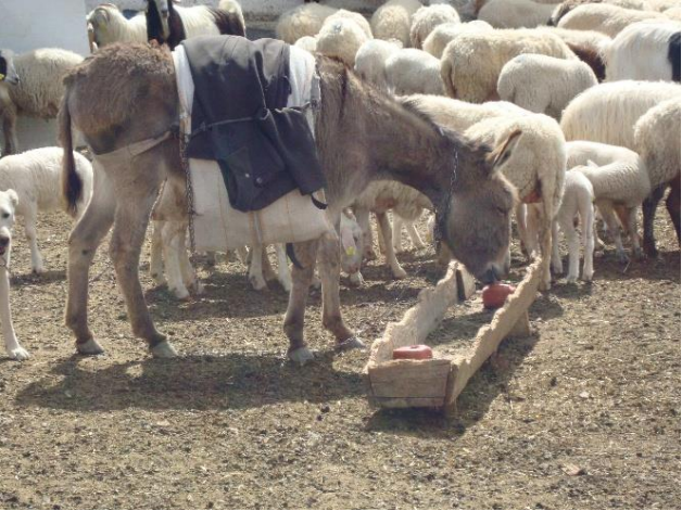
Based on a comprehensive analysis of the international literature and field data, the study, for the first time, systematizes the specific quantitative indicators of donkey nutritional needs based on their physiological condition and workload. It was found that a working donkey’s water requirement increased by 5-6 times, and its metabolic energy requirement increased by up to 2.5 times during an 8-hour workday compared to a resting period. For the first time, the detailed daily ration charts with a clear ratio of roughage and concentrated feed for donkeys weighing 100 and 200 kg and getting feed of various quality were developed and presented. The results confirm the high efficiency and safety of the proposed diets for maintaining health, productivity, and longevity of the animals. The study provides the science-based practical recommendations and forms a new knowledge base for proper feeding of donkeys in various keeping conditions.
This paper presents the nutritional requirements of donkeys, with a special focus on the needs for water, energy, proteins, minerals and vitamins. Donkeys, as working animals originating from arid regions, have specific physiological adaptations that affect their nutritional needs. The aim of this paper is to review and present scientifically based information on nutritional requirements, nutrients and recommendations for feeding donkeys based on current research in the world. The paper shows the needs for water, energy, proteins, minerals and vitamins, as well as methods of assessing food digestibility and availability. Special attention is paid to the variations in needs depending on the physiological state (growth, pregnancy, lactation), the intensity of work and the quality of available food. The importance of food digestibility and correctness of meal that meets all nutritional needs are also presented. The need for an adequate balance between rough and concentrated feed, as well as the potential danger of inadequate supplementation, are emphasized. Based on the available literature and field observations, practical instructions are given for proper feeding of donkeys in different breeding conditions, with the aim of preserving their working capacity, health and longevity.