Preview

Russian Journal of Veterinary Pathology

Advanced search

The “Russian Journal of Veterinary Pathology” is a peer-reviewed scientific and practical journal published since 2002 to enhance the level of the professional knowledge of the target audience on the latest achievements and prospects in the field of veterinary medicine of companion, agricultural and wild animals and to encourage the veterinary physicians to get the postgraduate education using the advanced freely available information space for modern scientific research and practical activity in the field of veterinary medicine. The results of the advanced fundamental and applied research are published as a priority; the scientific reviews in the field of theoretical and experimental veterinary medicine, biotechnology, as well as breeding, reintroduction, feeding and keeping of animals are presented; the cases from clinical practice that have scientific, factual, expedient significance are popularized; the materials on the history of veterinary medicine are included. Along with the discussion of the global trends in the above mentioned areas, attention is paid to the regional studies.

All articles are published in the Russian language and undergo a peer-review procedure.

The journal is included in the List of the peer-reviewed scientific publications (List of the Higher Attestation Commission under the Ministry of Science and Higher Education of the Russian Federation, 2nd category), where the main scientific results of dissertations for the degrees of Doctor and Candidate of Science in the following scientific specialties should be published:

1.5.17. Parasitology (biological sciences, veterinary sciences);

4.2.1. Animal pathology, morphology, physiology, pharmacology and toxicology (biological sciences, veterinary sciences);
4.2.2. Sanitation, hygiene, ecology, veterinary-sanitary examination and biosafety (biological sciences, veterinary sciences);

4.2.3. Infectious diseases and animal immunology (veterinary sciences, biological sciences);

4.2.4. Private animal husbandry, feeding, technologies for preparing feed and producing livestock products (biological sciences, agricultural sciences);

4.2.5. Breeding, selection, genetics and biotechnology of animals (biological sciences, agricultural sciences).

The target audience of the journal are the researchers and educators working in the field of veterinary science, the veterinary general practitioners, veterinary specialists, veterinary residents and postgraduate students.

The authors of the articles can be the practicing veterinary physicians, research associates and teachers of the educational and research institutions, postgraduate students and applicants for the academic degree of candidate and doctor of science, students of the veterinary science universities guided by the academic supervisors.

About the journal

The journal “Russian Journal of Veterinary Pathology” is registered by the Federal Service for Supervision of Communications, Information Technologies and Mass Communications on June 27, 2023 (Extract from the register of registered mass media ЭЛ № ФС 77–85552 – electronic edition).

All articles of the journal have DOI index registered in the CrossRef system.

Founder and publisher: Federal State Budgetary Educational Institution of Higher Education "Don State Technical University", Rostov-on-Don, Russian Federation, https://donstu.ru/

eISSN 2949-4826

Year of foundation: 2002.

Frequency: 4 issues per year (March 30, June 30, September 30, December 30).

Distribution: Russian Federation.

The journal “Russian Journal of Veterinary Pathology” accepts for publication original articles, studies, review papers, that have not been previously published.

Website: https://www.vetpat.ru/ 

Editor-in-Chief: Alexey М. Ermakov, Dr. Sci. (Biology), Professor (Rostov-on-Don, Russia).

Languages: Russian, English

Key characteristics: indexing, peer-reviewing.

Licensing history:

The journal uses International Creative Commons Attribution 4.0 (CC BY) license.

Current issue

Vol 25, No 2 (2026)
View or download the full issue PDF (Russian) | PDF

Animal pathology, morphology, physiology, pharmacology and toxicology

7-13 167
Abstract

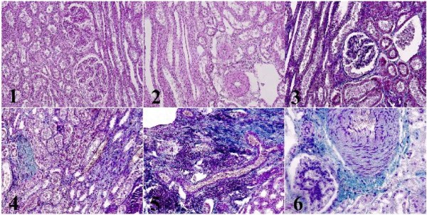
Introduction. Cow kidneys are exposed to the impact of industrial factors, which is manifested in development of the various nephropathies with systemic effects on organism. The kidneys are the main organs responsible for homeostasis by regulating osmolarity, mineral balance, and acid-base balance in organism through the excretion of water and inorganic electrolytes. Renal pathology often has latent character and is diagnosed postmortem. Significant destructive changes, including cell death, are often detected in lactating cows due to high metabolic load they undergo. Caspase pathway is one of the pathways in the complex apoptotic signaling cascade. Unfortunately, the expression of caspases in bovine kidneys has not been yet described in the literature. The aim of the study is to describe the relationship between the expression of caspase-3 and caspase-6 and the pathological changes in the kidneys of high-yielding cows.
Materials and Methods. A morphological study of renal samples from high-yielding cows (4–6 lactations, 7–8 years old, n=4) was conducted at the Ural Federal Agrarian Scientific Research Center, Ural Branch of the Russian Academy of Sciences in the period from 2023 to 2025. Standard histological techniques including hematoxylin and eosin staining and Mallory’s trichrome staining were used. Immunohistochemical analysis of the samples using antibodies to caspase-3, and -6 was performed.
Results. It was found that in the kidneys of 7–8-year-old cows there develop the destructive processes including renal failure, inflammatory reactions, and sclerotic and microcirculatory disorders. The observed conditions are associated with high expression of caspase-6 at the level of the renal tubules, which confirms damage to the renal parenchyma. Caspase-3 is expressed primarily in the renal tubules and in renal stroma elements, which indicates intensification of apoptotic processes across the entire organ.
Discussion and Conclusion. The destructive processes revealed in the kidneys of high-yielding cows are likely to be associated not only with the age but also with the high metabolic load imposed on the organism during lactation. The research findings substantiate the need for further study of pro-apoptotic factors due to their influence on the development of various pathologies in cattle.

14-24 168
Abstract

Introduction. Dopamine transporter gene knockout (DAT-KO) animal model is a valuable experimental tool for studying the pathophysiology of diseases associated with dopamine dysfunction, such as attention-deficit/hyperactivity disorder, schizophrenia, and Parkinson’s disease. The aim of the present review is to systematize the scientific data on using the DAT-KO rat models for studying dopaminergic dysfunction, epigenetic mechanisms, and gene therapy approaches.
Materials and Methods. Literary sources were searched for in PubMed, Scopus, Web of Science, and Google Scholar scientific citation databases. The original studies (n=49) on the structure and function of DAT, knockout models, epigenetic mechanisms, and gene therapy published from 1991 to 2025 in English language only were included into the review. The results have been presented in a PRISMA flow chart and in the illustrations.
Results. DAT knockout rat models exhibit a 5–7-fold increase of extracellular dopamine level, hyperactivity and structural changes in the basal ganglia, which indicates dopamine dysfunction. Homozygous knockout animals are found to completely lack functional DAT protein, whereas animals with heterozygous knockout retain approximately half of its function and exhibit intermediate phenotypes. Epigenetic regulation of SLC6A3 gene expression is mediated by DNA methylation, histone modifications (including H3K9/K14 acetylation and H3K27 methylation) and microRNA modifications. Moreover, the DAT promoter remains hypomethylated during postnatal ontogenesis in rats, resulting in its age-related expression increase. Gene therapy using viral vectors has demonstrated the potential to restore DAT function.
Discussion and Conclusion. The DAT-KO rat models reliably reproduce the key neurochemical and morphological features of dopaminergic dysfunction. However, the picture seems much incomplete due to the fragmentary character of data on the dynamics of epigenetic regulation of dopamine transporter expression during disease progression and insufficient evidence base on dose-dependent effects and long-term safety of gene therapy. The epigenetic mechanisms open the new tracks for biomarker search and personalization of therapy. Gene therapy using adeno-associated viral and lentiviral vectors demonstrates the potential to restore DAT function in the preclinical models, however, further clarification on dosedependent effects and minimization of the immune response is required. The integration of epigenetic markers into the clinical protocols, development of combinatory strategies, and validation of the DAT-KO models for comorbid conditions continue to be the promising tracks for further research.

25-33 133
Abstract

Introduction. Giving general anesthesia to animals is an important element of experimental research during the development and study of new veterinary drugs. Due to specific nature of general anesthesia and the need to use the potent sedatives and hypnotics, it has always been posing a risk to laboratory animals. In the frame of present-day veterinary research, the problem of proficient monitoring of anesthesia is extremely relevant, especially with regard to research involving laboratory rodents. The aim of the study is to develop and optimize laboratory monitoring methods of general anesthesia in animals during experimental studies of new veterinary drugs.
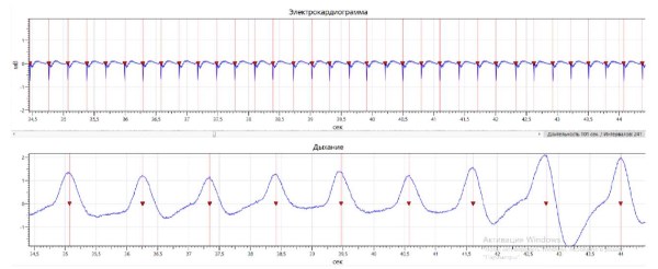
Materials and Methods. The study was conducted at the research laboratory of the Institute of Living Systems, DSTU, from January to May 2024. The experiment involved 20 Wistar rats of both sexes weighing 300–400 g that were subjected to inhalation anesthesia with isoflurane, using an RWD R520 anesthesia machine. The laboratory monitoring protocols were developed based on the electroencephalography (EEG), electrocardiography (ECG), respiratory rate, blood pressure, and body temperature parameters. The condition of animal organs and systems was assessed by means of biochemical blood and urine tests, as well as excretory function tests.
Results. An inhalation chamber connected to an anesthesia machine was designed for the study in rats. It allowed labor-atory monitoring of animals’ condition during inhalation anesthesia by electrophysiological and functional methods. A trial mode of the chamber using 2.0% isoflurane gas supply, at a flow rate of 2.0 l/min, and a withdrawal rate of 8.0 l/min had proved to be the optimal protocol for administering general anesthesia to laboratory rats. Electrophysiological, biochemical, and functional parameters demonstrated high tolerability of the anesthesia protocol used.
Discussion and Conclusion. The development proposed by the authors optimizes laboratory monitoring in animals during general anesthesia, contributes to improvement of quality of experimental research results, facilitates the work of re-searchers, and can be used during the development of new veterinary drugs.

34-43 140
Abstract

Introduction. The majority of pericardial diseases are accompanied by pathological accumulation of fluid in the pericardial cavity (effusion), as well as cardiac tamponade. Physical examination methods combined with transthoracic echocardiography are widely used to diagnose these conditions. An important task for veterinary specialists is to determine characteristic symptoms and specific echocardiographic signs of cardiac tamponade, which is considered to be a life-threatening condition. Cytological examination of pericardial fluid is an integral part in diagnosing the cause of the effusion. A comprehensive approach to diagnosis underpins the efficiency of treatment strategies. The existing studies poorly reveal the relationships between the clinical symptoms and echocardiographic signs, which could enable the diagnosis of disease severity and its prognosis, neither do they fully elucidate the cytological characteristics of effusions that could indicate the causes of effusion occurrence. The aim of the study is to establish the clinical, echocardiographic, and cytological relationships in dogs with pericardial pathologies accompanied by pericardial effusion.
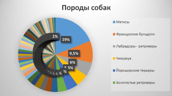
Materials and Methods. The study was conducted at the Department of Veterinary Surgery of Moscow State Academy of Veterinary Medicine and Biotechnology – MVA named after K. I. Skryabin and at the Veterinary Research Center “Biocontrol” in the period from 2018 to 2023. The objects under study were dogs of various breeds, ages, and sexes (n=200). The examination methodology included: collection of patient’s medical history, clinical examination, echocardiography, electrocardiography, chest radiography, ultrasonographic visualization of free fluid in the thoracic and abdominal cavities, pericardiocentesis, cytological examination of effusion, and histological examination of pathological material obtained during surgery.
Results. During the echocardiography, a ring-shaped anechoic space of varying diameter was revealed around the heart in all the studied dogs with pericardial effusion. Of 200 patients, cardiac tamponade was registered in 64 animals (32.0%), to whom pericardiocentesis was performed. For further determination of the causes of pericardial effusion, cytological examination of the obtained fluid was performed in 61 patients. According to cytomorphological characteristics, the following types of effusion were identified: transudate (1.6%), modified transudate (4.9%), septic exudate (1.6%), hemorrhagic effusion (which, in turn, was divided into effusion corresponding to: acute hemorrhage (4.9%), chronic hemorrhage (77.0%), hemorrhagic effusion with cytological signs of a tumor (9.8%)).
Discussion and Conclusion. In the presence of pericardial effusion, detection of the cardiac tamponade syndrome is of great clinical importance. We have established the most common echocardiographic signs of tamponade: caudal vena cava dilation and reduction of its collapsibility amplitude to less than 50%, as well as right atrial collapse. A relationship between the clinical symptoms (abdomen volume increase due to accumulation of ascitic fluid, progressing weakness, and exercise tolerance decrease) and echocardiographic signs of cardiac tamponade was revealed. Based on cytological examination results, the hemorrhagic type of pericardial effusion was identified as the most common one, concomitant to cardiac and pericardial tumors in older dogs of medium and large breeds.

44-53 144
Abstract

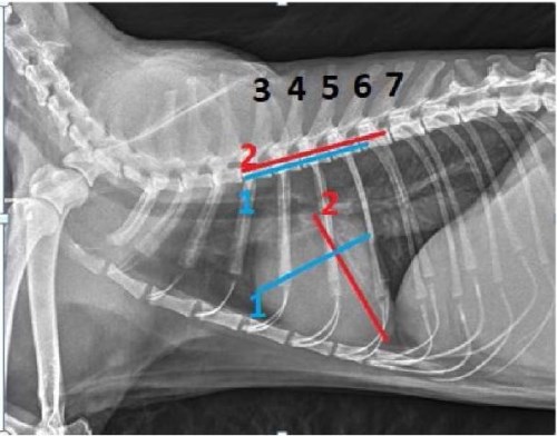
Introduction. Cardiac diseases are considered to be among the main causes of mortality in small domestic animals. Calculation of the vertebral heart score (VHS) is a method used to assess the cardiac silhouette enlargement on radiographs and predict the development of congestive heart failure. However, traditional manual procedure of VHS calculation is characterized by high inter-observer variability, subjectivity, and significant time-consumption. To improve the access to objective cardiac screening in every-day clinical practice, the authors has developed a desktop application for automating VHS calculation using the artificial intelligence (AI) technologies that unifies the assessment standard, minimizes human error, and accelerates processing of radiographs. The aim of the present pilot research is to conduct the first stage validation of the application and compare the diagnostic accuracy of two VHS calculation methods — semiautomated and expert-based.
Materials and Methods. A balanced sample of 30 right lateral chest radiographs of dogs and cats was selected for analysis as a result of simple random sampling. Preliminary VHS was calculated by manual measurements on the radiographs by the Buchanan & Bücheler method: two veterinarians specializing in visual diagnostics measured the short and long axes of the heart on the radiographs and then compared their dimensions against the spinal vertebrae. The semi-automated calculation of VHS was performed by the author-developed “Desktop application for automation of the vertebral heart score (VHS) calculation based on radiographs” using a convolutional neural network. App validation implied a comparison of the results of the two methods of VHS calculation, taking into account the intraclass correlation coefficient (ICC).
Results. The “Desktop application for automation of the vertebral heart score (VHS) calculation based on radiographs” has proved to perform a highly accurate and replicable automated calculation of VHS on lateral chest radiographs of dogs and cats. It also has demonstrated statistically high agreement with the calculations obtained by veterinarians specializing in visual diagnostics (ICC≥0.97), along with less time required for calculation.
Discussion and Conclusion. The first stage of app validation has demonstrated an excellent agreement between the AI and two specialists’ results, therefore, the proposed approach can be deemed a future-oriented tool for general practitioners. The present pilot research has several limitations, i.e. it was conducted on a relatively small sample of animals, which may be insufficient for evaluation of app performance in narrow subgroups (e.g., in specific breeds or in severe cardiomegaly cases). Further studies in larger, more heterogeneous cohorts, including patients with severe cardiomegaly and anatomical abnormalities, are required to confirm clinical significance of the results.

54-60 147
Abstract

Introduction. At the contemporary stage of mammary tumor studies, the microenvironment is considered a key factor contributing to neoplasia formation and metastasis. Therefore, the greatest attention is paid to the cellular composition of the mammary tumor microenvironment, in which the most widespread elements are the stromal cancer-associated fibroblasts (CAFs). However, in veterinary oncology, stromal markers of malignant mammary tumors are insufficiently studied. The aim of the present study is to investigate the expression of tumor marker proteins, i.e. vimentin, α-SMA, and CD34, in the stroma of the malignant mammary tumor microenvironments in dogs.

Materials and Methods. The objects of the study that was conducted in the period from 2022 to 2024 were the female dogs of various breeds and ages (n=26). The materials studied were the mammary tumors. A comprehensive range of histological and immunohistochemical studies was conducted in compliance with the standard techniques.
Results. It has been established that in different histological types and degrees of canine mammary tumor malignancy, the fibroblastic differentiation lineage cells demonstrate a high expression of vimentin and α-SMA and a low expression of CD34, which indicates their differentiation into CAFs.
Discussion and Conclusion. The obtained results can be used as basic applied data in the study of carcinogenesis in veterinary oncology.

Infectious diseases and animal immunology

61-67 134
Abstract

Introduction. Active development of biotechnologies and genetic engineering techniques broadens the horizons for engineering the probiotic bacterial strains with tailored functional properties, including the strains based on bacteria of the genus Lactobacillus that are traditionally considered safe microorganisms. Genetic modification enhances their antagonist activity, resistance to adverse environmental factors, and boosts metabolic efficiency. However, bacterial genome editing may entail risk of introducing the undesirable traits, including cytotoxicity and expression of virulence factors. Safety concerns are particularly relevant in the frame of veterinary practice, where probiotics are widely used to correct the gastrointestinal microbiocenosis in agricultural animals and improve their productivity. The aim of the study is to conduct a comprehensive analysis of cytotoxicity of the genetically modified Lactobacillus plantarum 8PAЗ (B-11007) strain including identification and evaluation of the virulence factors responsible for inducing cell death and disruption of cellular functions.
Materials and Methods. A genetically modified Lactobacillus plantarum 8PAЗ (B-11007) strain was the object of the study conducted in the period from January to February 2026. A comprehensive in vitro analysis of its cytotoxicity included: assessment of the cytopathic effect in intestinal cell culture, the effect on the viability and metabolic activity of enterocytes, and the state of intestinal epithelial barrier function. Additionally, the analysis of potential virulence factors was performed, along with the study of the mechanisms of cell death, including signs of apoptosis and necrosis.
Results. The recombinant Lactobacillus plantarum 8PAЗ (B-11007) strain has demonstrated a high biological safety and pronounced biological activity, which enhanced the intestinal epithelial barrier function by 15.2%, the metabolic activity of cells by 8.7% and simultaneously reduced early apoptosis of enterocytes by 31.1% (p<0.05), showing no signs of cytotoxic action or activation of caspase-dependent cell death.
Discussion and Conclusion. The conducted comprehensive analysis allows making the unequivocal conclusion about genetically modified L. plantarum 8PAЗ (B-11007) strain exhibiting no cytotoxic activity in vitro. Moreover, by certain properties (cellular metabolic activity, barrier function), it exceeds the original native strain, which makes it a promising candidate for further preclinical and clinical trials as a potential probiotic agent.

Announcements

More Announcements...


Creative Commons License
This work is licensed under a Creative Commons Attribution 4.0 License.